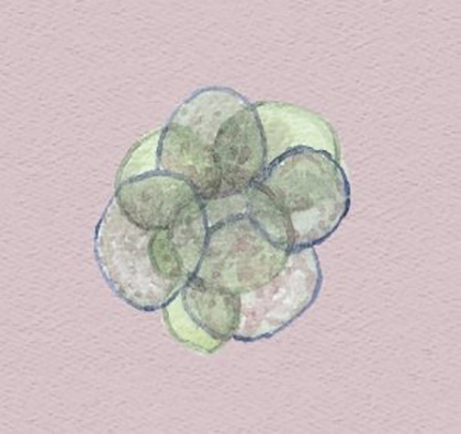
Fragment removal
From the very moment when sperm fertilises an ova, a new embryo starts developing and a large number of cell divisions take place. This embryo development is observed in an in vitro fertilisation laboratory up until the blastocyst stage (day 5 or 6 of development). Sometimes during the cell division process, fragments of the embryo become isolated between cells that have developed correctly. These fragments come from embryo cell remains and can stop the embryo from developing correctly. One of the negative impacts consists of issues reaching the blastocyst stage and the posterior impact on implantation in the uterus. In fact, embryo fragmentation is one of the most significant characteristics used to determine embryo quality.
Image of a human embryo on day 3 following fertilisation using an electronic microscope. On the left, an embryo in 8-cell stage with almost no fragmentation. On the right, an embryo in 8-cell stage with approximately 25% fragmentation (A. Jurisicova, 1996).
Fragment removal is an assisted reproduction technique used to eliminate cell fragments. First of all, the outer-most layer of the embryo (zona pellucida) is perforated and then the largest possible number of cell fragments are aspirated.
Whilst recent research suggests a link between embryo cell death and increased fragmentation (HJ. Chi, 2011), there are a number of research studies that indicate that removing these fragments is not effective and does not improve implantation of the embryo in the uterus. The technique is invasive and handling the embryo can affect posterior development and, for this reason, using the technique is highly controversial.
