What is the impact of vitrification on biopsied blastocysts?
Selecting healthy and viable embryos is increasingly important in the field of assisted reproduction. The selection is made using pre-implantation genetic diagnosis (PGD) which consists of analysing some of the cells in the embryo and determining if it is healthy before it is transferred to the patient.
Embryos can currently be retained in culture until day 5 to 7 of development when they reach blastocyst stage. During this period, the embryo can be transferred or vitrified for transfer in the future.
Why do we freeze blastocysts?
Embryos are vitrified when:
- Following a cycle, several embryos from a single cohort are still available because standard practice means transferring a single embryo and vitrifying the remainder.
- There is a medical recommendation indicating that transfer should be deferred because of a potential risk of hyperstimulation.
- When it is not possible to perform embryo transfer on the selected day due to the patient’s personal circumstances.
- We perform biopsies on blastocysts and we have to wait for the genetics results before transfer.
When is performing a biopsy on a blastocyst most effective?
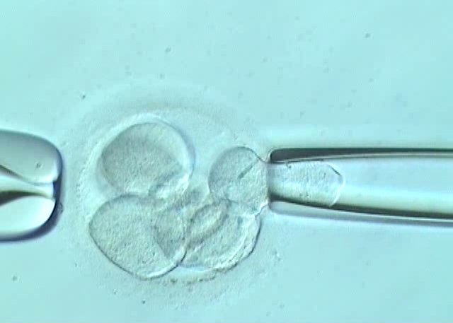
When there is a PGD recommendation, we perform assisted hatching in the assisted reproduction laboratory with embryos on day 3 of development. This consists of using laser to make a small opening in the zona pellucida of the embryo. It helps the embryo to begin extruding cells from the trophectoderm once it has reached blastocyst stage. The embryo biopsy is completed during this phase, after days 5 or 6 of development. It consists of removing cells from the trophectoderm using microhandling equipment and laser. Between 4 and 6 cells are generally retrieved and these are taken to the laboratory at IB Biotech so that genetic analysis can be performed on them. At this point, the blastocyst needs to be vitrified and we must wait for the results of the genetic diagnosis before transfer.
At Instituto Bernabeu…
we have an excellent embryo vitrification programme with survival rates in non-biopsied embryos reaching 95%. So far, we have observed 100% survival rates in biopsied embryos. This leads us to believe that the fact that these embryos have been subjected to a biopsy may give them an advantage over others. The balance process using cryprotectants delivers this increase in survival rates due to more direct exposure of the embryo to the balance medium because the zona pellucida in these embryos has been perforated and they are also either fully or partially exposed.
This high survival rate means that we can firmly believe in the blastocyst biopsy strategy followed by cryopreservation, whether this be in order to screen all the chromosomes or for diagnosing a monogenic disorder.
Miguel Herreros, a biologist at Instituto Bernabeu.
