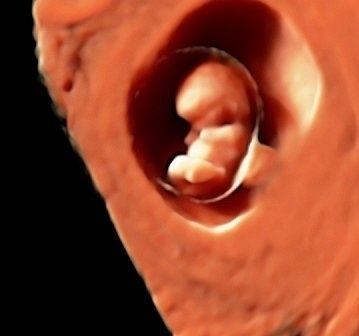
Recurrent pregnancy loss. Embryo implantation failure.
Perhaps one of the most devastating feelings that both doctors and patients have to endure during in vitro fertilisation (IVF) treatment is finding out that pregnancy has not been achieved following several attempts, or that a pregnancy has been interrupted early due to recurrent pregnancy loss. In other words, implantation failure.
Given that this situation generates a great deal of frustration amongst patients, it can lead to empirical ‘courses of treatment’ promising ‘miracle’ solutions.
Dealing successfully with cases of this kind is one of the biggest challenges currently faced by specialists in human reproduction.
Let us take a look, then, at how we successfully deal with this issue.
Embryo implantation is an incredibly complex process that requires a combination of a suitable embryo and a receptive endometrium, as well as good communication between the two.
We must not forget, of course, that only one in every four embryos implant naturally, so this is the biggest challenge we face and a great unknown.
Both maternal and paternal embryo factors cause implantation failure. Therefore, it is essential that a detailed analysis of them all is carried out before beginning a new course of treatment that would otherwise necessarily just be a source of additional, and often unbearable, frustration for patients.
The analysis should be accompanied by careful evaluation of all courses of treatment carried out so far.
By way of breaking things down into simple terms, let us take into account that there are three leading characters: the mother, the father and the embryo.
The MATERNAL contribution to this situation is the most complex one. There may be issues in the uterus such as malformations, tumours, scars, adhesions, infections or inflammation that affect uterine receptivity. This calls for careful evaluation using a diagnostic hysteroscopy, cultures from the uterus and, sometimes, an analysis of the area where the embryo implants using an endometrial biopsy.
Issues in the ovary are the second factor that need to be analysed, including observation of ovarian response, the number and quality of oocytes retrieved and their evolution in the cycles carried out so far.
We will take another look at this when we talk about the embryo. It is also essential to rule out infections near to the uterus such as hydrosalpinx and the consequences of earlier infections in the fallopian tubes.
Last of all, we look at immunological or blood clotting defects that are an impediment to implantation. Analysis is carried out through a blood sample to detect the presence of antibodies that negatively affect implantation and abnormalities in coagulation that lead to small microthrombi affecting the normal blood flow for feeding the embryo in its early stages of development.
Chromosome abnormalities need to be analysed in both partners. A karyotype, which is an analysis of chromosomes (the structures that transport our inherited genetic material), provides us with very useful information that sometimes needs to be backed up with more specific tests that we will take a look at later on.
The second leading character is the FATHER. Apart from the indicated karyotype, a basic semen analysis, providing an understanding of the percentage of spermatozoa with fragmented DNA and their chromosome make-up using TUNEL and FISH tests, enables us to get into minute detail on the paternal contribution to the future embryo.
Finally, we have evaluation of the third patient: the EMBRYO.
Unfortunately, the information we are currently able to retrieve is limited.
However, in a detailed evaluation of the cycles previously carried out – number of oocytes obtained, fertilisation rate, techniques used, technological and human resources used in those courses of treatment, a detailed evaluation of embryo development during the period in the laboratory and up until the day on which they are transferred – the characteristics that they do have are essential to providing an accurate prognosis.
Once all this has been completed, we should be able to suggest solutions and provide the couple with a prognosis. Personalised treatment is the best way forward in most cases.
Whilst we must not forget that experience indicates that couples who have gone through 3 early pregnancy losses have up to a 70% change of achieving a successful pregnancy on their own without having to go through treatment of any kind, several techniques do improve results.
First of all, correcting the issues that were diagnosed during the analysis. Treating maternal and paternal factors will ensure that the condition of embryo and the uterus improve considerably, increasing the chances of correct implantation and embryo development.
Additionally, an analysis of the embryo’s chromosomal make-up with pre-implantation genetic diagnosis (PGD), or supporting hatching when the time has come for the embryo to implant in the mother’s uterus.
In some cases that cannot be treated, we have to turn to oocyte or sperm donation. These options have very high success rates.
The outlook is that molecular biology provides hope for analysis and treatment in couples of this kind. Nowadays, research work aimed at understanding embryo metabolism enables us to get a good understanding of early needs and means of functioning.
Instituto Bernabeu is a leading entity in embryo metabolomics.
Step by step, the results are improving prognoses and we are able to provide increasingly more help.
We have recently acquired and implemented a new state-of-the-art technique called array CGH that is far more sensitive and efficient than the conventional karyotype (it enables identification of duplications or absences of small chromosome regions that are not detected in the karyotype). That is, a level of diagnosis that is better 10-fold. This technique is a huge step forward for cases of sterility with an unknown cause and of repeated implantation failure (get to know more about array CGH).
Dr Rafael Bernabeu, Medical Director at Instituto Bernabeu.
